


Ultrasound
Ultrasound is a painless, non-invasive, diagnostic test that produces images without the use of radiation it produce images of organs and soft tissue
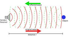
a non-invasive technique that is used to generate real-time images of the body’s internal structures Through a series of high-frequency sound waves,
ultrasound technology allows for the visualization of fetal development, blood flow, and internal structures, making it effective for both diagnostic
and pathologic assessment. It is a valuable resource for tracking the health of a patients. Ultrasound is also used to view organs such as the liver,
uterus, ovaries, thyroid, and aorta. It is also used to help evaluate blood clots or the narrowing of vessels.
What to expect?
The examination is performed by specialized technicians (medical imaging technologists) or by a radiologist. It takes place in a room containing a
bed and an ultrasonic device.
The examination is painless:
The sounds emitted by the device are inaudible.
It is fast.
It requires no preparation.
Gel will be applied on your skin around the imaging area:
You may find it a bit cold when applied.
You will be asked to put on a gown or to bare the imaging area.
You will lie down on the bed and the probe will be applied to the surface of your skin.
Types of Ultrasound Scans
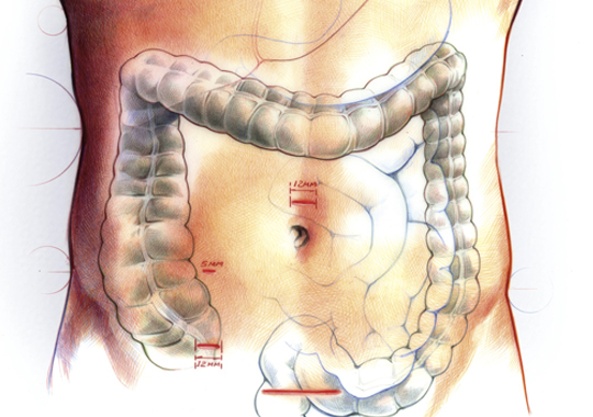
Abdominal/Pelvic Ultrasound Combined
Gallbladder, Abdominal
Pelvic, Renal - scans of kidneys, cysts, stones or the bladder
Abdominal Ultrasound
Pelvic/Gyne Ultrasound
Obstetrical Ultrasound
Pelvic Transabdominal & Transvaginal
Arterial Doppler lower and upper extremity
Venous Doppler lower and upper extremity
Biophysical Profile
Renal Ultrasound
Thyroid
Copyrights © All Rights Reserved Toronto Liver Centre